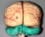
Major Parts of the Brain
- Miroslav Czadek
- Aug 24, 2024
- 5 min read
/** upadated 24-08-24 **/
I. Cerebrum
The cerebrum is the largest part of the brain, comprising two cerebral hemispheres. It is responsible for most higher brain functions, including sensory perception, motor control, language, thought, memory, emotions, and decision-making processes. The cerebrum is divided into several regions, each with specialized functions:
1. Frontal Lobe: The anterior part of the cerebral cortex, located behind the forehead. It is crucial for executive functions such as decision-making, problem-solving, planning, voluntary motor activity, and controlling behavior and emotions. It also contains Broca’s area, which is associated with speech production.

2. Parietal Lobe: Positioned at the upper back area of the brain, just behind the frontal lobe. This lobe processes sensory information from the body, including touch, temperature, and pain, and integrates spatial information to help with navigation and movement. It also houses Wernicke’s area, which is involved in language comprehension.

3. Temporal Lobe: Located on the sides of the brain, near the temples, beneath the frontal and parietal lobes. The temporal lobe is essential for processing auditory information, language comprehension, and memory formation. It also plays a role in emotion and recognizing faces.

4. Occipital Lobe: The posterior part of the brain, located at the back of the skull. The occipital lobe is primarily responsible for visual processing, including interpreting visual stimuli and identifying objects.
5. Insula: A small region of the cerebral cortex located deep within the lateral sulcus. The insula is involved in diverse functions, such as emotion, self-awareness, homeostasis, and interoceptive awareness (sensing the internal state of the body).

6. Hippocampus: A seahorse-shaped structure located in the medial temporal lobe. It is crucial for memory formation, particularly in the transition of short-term memory to long-term memory, and is involved in spatial navigation.

7. Lateral Ventricles: Two large, C-shaped cavities within each hemisphere of the cerebrum, filled with cerebrospinal fluid. They protect the brain by cushioning it, removing waste products, and circulating cerebrospinal fluid which provides nutrients to the brain.

8. Corpus Callosum: A thick bundle of nerve fibers that connects the left and right cerebral hemispheres. It enables communication between the two hemispheres, allowing for coordinated brain function and the integration of motor, sensory, and cognitive tasks.

9. Fornix: An arched bundle of fibers that carries signals from the hippocampus to other parts of the brain, including the mammillary bodies. It is involved in memory processing and recall, particularly in transmitting information within the limbic system, which is associated with emotions and memory.
II. Cerebellum
The cerebellum is located under the cerebrum at the back of the brain, consisting of two hemispheres and a central vermis. It is essential for motor control, balance, and coordination. The cerebellum fine-tunes voluntary movements and helps maintain posture, balance, and motor learning.
10. Vermis: The midline structure that connects the two hemispheres of the cerebellum. It plays a critical role in posture and locomotion by controlling the axial (trunk) muscles.

11. Hemispheres: The lateral portions of the cerebellum on either side of the vermis. These hemispheres coordinate voluntary movements, such as the fine motor movements of the limbs, and contribute to motor learning and timing.

III. Diencephalon
The diencephalon is located between the cerebrum and the brainstem and consists of structures like the thalamus, hypothalamus, and other vital components. It acts as a relay center for sensory information and controls various autonomic functions. The diencephalon is crucial for homeostasis, sensory perception, and endocrine function.
12. Thalamus: A large, dual-lobed mass of gray matter located deep in the brain. It acts as the brain’s relay station for sensory and motor signals, sending information to the cerebral cortex. The thalamus also plays a key role in regulating consciousness, sleep, and alertness.
13. Hypothalamus: A small but vital structure located below the thalamus. It controls many autonomic functions, including temperature regulation, hunger, thirst, and circadian rhythms. The hypothalamus also links the nervous system to the endocrine system via the pituitary gland, influencing hormone release.

14. Hypophysis (Pituitary Gland): A small, pea-sized gland located at the base of the brain, connected to the hypothalamus. Known as the “master gland,” it regulates various physiological processes by releasing hormones that influence other endocrine glands, controlling growth, metabolism, and reproductive functions.
15. Optic Nerve: The second cranial nerve, extending from the retina to the brain. It is essential for vision, transmitting visual information from the eyes to the brain, where it is processed to form images.
16. Pineal Body: A small, pinecone-shaped endocrine gland located near the center of the brain. It produces melatonin, a hormone that regulates sleep-wake cycles and circadian rhythms.

17. Medial Geniculate Body: Part of the auditory thalamus, it serves as a relay station in the auditory pathway, processing sound information and transmitting it to the auditory cortex.
18. Lateral Geniculate Body: Part of the visual thalamus, it plays a crucial role in visual processing, receiving inputs from the retina and sending them to the visual cortex for interpretation.
IV. Mesencephalon (Midbrain)
The mesencephalon, or midbrain, is a portion of the brainstem located between the diencephalon and the pons. It plays an important role in vision, hearing, motor control, sleep/wake cycles, arousal (alertness), and temperature regulation.
19. Superior Colliculi: Paired structures located on the dorsal surface of the midbrain, involved in visual processing, particularly in coordinating eye movements and visual reflexes, such as tracking moving objects.

20. Inferior Colliculi: Paired structures found below the superior colliculi, critical for auditory processing. The inferior colliculi relay sound information from the ear to the auditory cortex and are involved in auditory reflexes.
21. Trochlear Nerve (IV): The fourth cranial nerve, emerging from the dorsal aspect of the brainstem. It innervates the superior oblique muscle of the eye, enabling downward and inward eye movements.
22. Oculomotor Nerve (III): The third cranial nerve, originating in the midbrain. It controls most of the eye’s movements, including the raising of the eyelid, constriction of the pupil, and the ability to focus on objects.

V. Pons
The pons is a broad, horseshoe-shaped structure located on the brainstem, between the midbrain and the medulla oblongata. It serves as a communication and coordination center between the two hemispheres of the brain. The pons plays a key role in regulating breathing, sleep, and relaying information between the cerebrum and cerebellum.
23. Trigeminal Nerve (V): The fifth cranial nerve, with three major branches, is responsible for sensation in the face (touch, pain, temperature) and motor functions such as biting and chewing.
24. Abducent Nerve (VI): The sixth cranial nerve, originating from the pons, controls the lateral rectus muscle of the eye, responsible for moving the eye outward (abduction).
25. Facial Nerve (VII) and Intermediate Nerve: The facial nerve controls muscles for facial expressions, while the intermediate nerve is involved in taste sensation and controlling glands like the salivary glands.

26. Vestibulocochlear Nerve (VIII): The eighth cranial nerve is essential for hearing and balance, transmitting sound and equilibrium information from the inner ear to the brain.
VI. Medulla Oblongata
The medulla oblongata is the lowest part of the brainstem, connecting the brain to the spinal cord. It regulates vital autonomic functions such as heart rate, blood pressure, breathing, and reflexes like swallowing and coughing.
27. Pyramid: Two ridges on the anterior surface of the medulla oblongata, containing motor fibers that transmit signals from the brain to the spinal cord, facilitating voluntary motor control.
28. Olive: An oval-shaped prominence on the medulla, adjacent to the pyramids, involved in motor learning and coordination, as well as relaying sensory information to the cerebellum.
29. Glossopharyngeal Nerve (IX): The ninth cranial nerve provides taste sensation from the posterior third of the tongue, contributes to swallowing, and controls the secretion of saliva.
30. Vagus Nerve (X): The tenth cranial nerve controls a wide range of parasympathetic functions, including heart rate, digestive tract activity, and reflexes like coughing.
31. Accessory Nerve (XI): The eleventh cranial nerve controls muscles involved in head movement, specifically the sternocleidomastoid and trapezius muscles.
32. Hypoglossal Nerve (XII): The twelfth cranial nerve controls the muscles of the tongue, which are essential for speech, swallowing, and food manipulation.
